What Part of the Brain Areas Causes Autism
Discover what part of the brain causes autism and explore its impact on development and behavior.

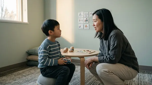
The neurologist used a phrase you did not quite expect, something about "atypical brain connectivity," and you have been turning it over in your head ever since. The visit ended with a recommendation for therapy and a follow-up scheduled six months out, but the question that stuck with you walking back to the car was simpler than what they explained: which part of the brain is actually doing this? And does the answer change anything about what you should do next?
The honest answer is that autism is not caused by one region of the brain in the way a stroke might affect one specific area. Decades of research, drawing on brain imaging, genetic studies, and developmental neuroscience, point to a pattern of differences across multiple regions and the connections between them, shaped by both genetic and environmental factors [1]. The rest of this article walks through what we know about brain development, structure, and connectivity in autism, and what the science means for parents who are weighing what kind of support will help most.
Brain Development in Autism
Understanding brain development in children with Autism Spectrum Disorder (ASD) involves examining the interplay between genetic and environmental factors, along with the structural differences researchers have identified in the brains of people with autism.
Genetic Influences on Brain Development
Genetic factors play a substantial role in autism. Changes in certain genes or the genome can increase the likelihood that a child will develop autism. Some of these genetic changes are inherited from parents, which means autism often shows up in family patterns. A meta-analysis of twin studies suggests that 60 to 90 percent of the risk for autism stems from genetic factors [3]. This strong genetic component highlights the importance of considering inherited traits when researching autism.
| Study Type | Percentage of Genetic Influence |
| Twin Studies | 60 to 90% |
Environmental Factors in Brain Development
Environmental factors also affect brain development. Factors such as prenatal exposure to certain medications or toxins, birth complications, and other early-life stresses can contribute to differences in brain development. These factors, in combination with genetic predispositions, may shape how autism eventually presents. Researchers continue to work on identifying the specific environmental conditions that interact with genetic factors in autism.
Brain Structural and Growth Differences
Many studies indicate that children with Autism Spectrum Disorder show various structural differences in their brains. These differences include changes in total brain volume, specific regional structures, and the cortex. Research has shown that children with autism often display accelerated growth in total brain volume, particularly during early childhood, with the pattern sometimes stabilizing or normalizing as they get older.
| Age Group | Brain Volume in ASD (Comparison) |
| Early Childhood | Often enlarged compared to typically developing peers |
| Adolescence | Stabilization observed |
| Adulthood | Variable; some have normalized |
Specific regions, such as the right inferior frontal gyrus, have been implicated in differences related to language development and social interaction. Changes in these areas may affect a child's ability to understand nuanced communication, such as irony or implied meaning. Alterations in white matter, especially within the corpus callosum (the band of fibers that connects the two hemispheres), suggest that structural changes are closely linked to autism characteristics.
| Type of Difference | Description |
| Total Brain Volume | Accelerated growth observed in early childhood |
| Regional Brain Structure | Differences in areas such as the right inferior frontal gyrus |
| White Matter | Alterations in the corpus callosum linked to autism traits |
These insights into brain development help illustrate why the question "what part of the brain causes autism" does not have a single clean answer. It is a pattern of structural and developmental differences, shaped by a mix of genetic and environmental factors, that together produce the wide range of autism presentations we see in real children.
Neuroimaging Techniques in Autism
Neuroimaging techniques have significantly advanced understanding of brain function and structure in autism. Two prominent methods used in this research are EEG (Electroencephalogram) and fMRI (Functional Magnetic Resonance Imaging).
EEG (Electroencephalogram) Studies
EEG measures the electrical activity of the brain through electrodes placed on the scalp. This method can gather data from tens to hundreds of electrodes positioned at various locations on the scalp. EEG offers insights into both spontaneous and event-related brain activity, which makes it valuable for cognitive neuroscience research.
Some key features of EEG include:
| Feature | Description |
| Temporal Resolution | High (milliseconds) |
| Main Function | Measures neuronal responses |
| Application | Understanding brain activities and connectivity patterns |
EEG's high temporal resolution lets researchers track the dynamics of brain activity as it unfolds, which is essential for studying how the brain responds to specific stimuli moment to moment.
fMRI (Functional Magnetic Resonance Imaging) Studies
fMRI uses magnetic resonance imaging to observe brain activity by measuring changes associated with blood flow. Unlike EEG, fMRI provides high spatial resolution and comprehensive brain coverage, although it has slower temporal resolution because the hemodynamic response itself takes a few seconds.
Key characteristics of fMRI include:
| Feature | Description |
| Spatial Resolution | High (covering entire brain) |
| Temporal Resolution | Lower (compared to EEG) |
| Main Function | Measures brain activity through blood flow |
Task-based fMRI studies have revealed atypical activation in areas critical for social communication and restricted repetitive behaviors (RRBs) in people with ASD. Key brain regions identified include:
| Brain Region | Role in ASD Symptoms |
| Inferior Frontal Gyrus | Language processing |
| Superior Temporal Sulcus | Face recognition |
| Frontoparietal Cortex | Executive functions |
| Amygdala | Emotional regulation |
These findings indicate that some of the patterns seen in autism may also overlap with other conditions like obsessive-compulsive disorder and generalized anxiety, which is one reason why diagnosis relies on behavioral assessment rather than imaging.
Combining EEG and fMRI data enhances understanding of brain activity by drawing on the strengths of each. This integrated approach continues to deepen what we know about how the autistic brain processes information.
Brainstem Involvement in Autism
Understanding the role of the brainstem in Autism Spectrum Disorder is important, since this region is involved in many autonomic functions and basic neurophysiological processes. Research suggests that differences in early brainstem development may be associated with how autism shows up later.
Early Brainstem Development
The early stages of brainstem development are critical. Environmental factors that disrupt this phase have been linked to an increased incidence of ASD. Certain teratogenic exposures, such as valproic acid and thalidomide during early pregnancy, are associated with a higher prevalence of autism in children. Disturbances during the period of rapid brainstem development may correlate with the emergence of autism characteristics.
Pre-term infants often face challenges due to underdeveloped brainstems. They can show irregular autonomic functions, such as differences in temperature regulation and heart rate variability. Some of these patterns overlap with what is observed in children with ASD, which suggests a possible developmental connection between brainstem maturation and autism-related characteristics.
Brainstem Differences in ASD
Research using Auditory Brainstem Response (ABR) studies has shown atypical auditory function in both pre-term infants and children diagnosed with ASD. Atypical ABR findings have been associated with differences in attention and social engagement in children with autism, suggesting that brainstem neurophysiology may play a role in core characteristics of the disorder.
Histological examinations of the brainstem in people with ASD have revealed notable morphological differences. Notable findings include:
| Brainstem Substructure | Observed Changes |
| Arcuate Nucleus | Atypical connectivity affecting sympathetic and parasympathetic tone |
| Inferior Olivary Nucleus | Imbalances linked to motor and sensorimotor differences |
| Superior Olivary Complex | Atypical structures associated with auditory processing |
These findings point toward specific brainstem differences that may contribute to the developmental picture of ASD. The variety of patterns observed signals the need for further investigation into how brainstem differences relate to autism's behavioral characteristics.
In our practice, parents most often start asking these neuroscience questions at one of two moments: right after a diagnosis, when they want to understand what is happening biologically, or about a year into therapy, when they have watched their child make progress and want to understand why some skills come more easily than others. The science is reassuring on both counts: brain differences are not a verdict. They describe starting conditions, not outcomes. Positive reinforcement and structured teaching actively shape how the brain organizes new skills, particularly during the years of greatest plasticity.
Brain Connectivity in Autism
Understanding brain connectivity is central to deciphering what is different about the autistic brain. This section focuses on hypo- and hyper-connectivity, and on functional and structural connectivity in people with Autism Spectrum Disorder.
Hypo- and Hyper-Connectivity
Research shows that people with ASD exhibit both hypo-connectivity (reduced communication between certain areas) and hyper-connectivity (enhanced connectivity in other areas). The distinction matters because it helps explain the wide range of ways autism shows up.
| Type of Connectivity | Examples of Affected Regions | Implications |
| Hypo-Connectivity | Language processing areas, face recognition regions, visuomotor coordination | Differences in executive function and challenges in social communication |
| Hyper-Connectivity | Salience, default mode, frontotemporal, motor, and visual networks | Alters perception and response to social cues |
Resting-state functional MRI studies have shown that adolescents and adults with ASD often exhibit hypo-connectivity in regions such as the default mode network (DMN) and the right posterior superior temporal sulcus, which affects how language and social interactions are processed.
Functional and Structural Connectivity
Both functional and structural connectivity in the brains of people with ASD can be measured to reveal patterns that correlate with autism characteristics.
Functional Connectivity examines how active different regions of the brain are when a person is at rest or engaged in a specific task. In ASD, task-based fMRI studies have identified differences in activation in areas associated with social communication and restricted repetitive behaviors.
Structural Connectivity involves measuring the physical connections between brain regions, often using methods such as diffusion tensor imaging. Structural studies have highlighted weaker connections in regions responsible for speech motor planning, which may contribute to challenges in expressive communication for some people with ASD.
Key findings include:
| Measurement Type | Findings |
| Functional Connectivity | Atypical activation in the inferior frontal gyrus, superior temporal sulcus, and amygdala linked to social differences and RRBs [2] |
| Structural Connectivity | Weaker connections in speech production networks, indicating differences in motor planning for communication [2] |
Connectivity findings are particularly relevant to therapy, because connectivity is one of the things that changes with repeated, structured practice. When a child learns a new skill, neural pathways strengthen; when that skill is practiced across multiple settings, those pathways generalize. This is exactly what we are doing when we work on how to foster generalization of learned skills across home, school, and community settings.
Implications for ASD Symptoms
Understanding how autism affects a child involves looking at the implications for social communication and cognitive functioning. Differences in brain structure and connectivity show up in the kinds of skills children find harder, and in the kinds of supports that help.
Social Communication Differences
Children with ASD often face significant challenges in social communication. These can show up as differences in social interaction, delayed or atypical language development, and difficulty perceiving emotional cues that other children pick up automatically. Research indicates that there is atypical activation in several brain regions during tasks associated with emotional processing. Key areas include:
| Brain Region | Associated Function |
| Amygdala | Emotion recognition and processing |
| Superior Temporal Sulcus | Interpretation of social cues |
| Inferior Frontal Gyrus | Language and social interaction |
Differences in these areas are linked to challenges in theory of mind (the ability to understand others' thoughts and feelings) and in real-time monitoring of social responses [2]. These cognitive elements are foundational for building relationships and navigating everyday communication.
Cognitive Differences
Cognitive profiles in children with ASD vary widely, but commonly include differences in working memory, language processing, and executive function. Task-based fMRI studies have revealed atypical activation patterns in brain regions responsible for these functions:
| Cognitive Function | Brain Region | Activation Findings |
| Language Processing | Inferior Frontal Gyrus | Imbalanced activation during language tasks |
| Face Recognition | Superior Temporal Sulcus | Atypical responses to emotional faces |
| Executive Function | Frontoparietal Cortex | Dysregulated activity affecting decision-making and tasks |
Children with ASD often show hypo-connectivity in areas relevant to language processing, facial recognition, and executive functioning, which suggests weaker neural connections in these networks [2]. This affects social communication and many of the daily cognitive tasks that depend on coordinating across regions.
The practical takeaway for parents: brain biology helps explain why some skills are harder. It does not predict where the ceiling is. The years of greatest brain plasticity are also the years when high-quality, individualized intervention has the most impact, which is why starting your child's ABA journey with early intervention matters so much.
Why Families Choose Mastermind Behavior
Mastermind Behavior is a BCBA-owned in-home ABA therapy provider serving families across New Jersey, Georgia, and North Carolina. Every child's program starts with a Board Certified Behavior Analyst (BCBA), who reviews evaluations, conducts our own assessment, and designs an individualized plan. Behavior Technicians (BTs) carry out the daily teaching sessions in your living room, kitchen, and backyard, the real environments where new skills need to take hold. Our parent training coaches help you fold the same approaches into bedtime, morning routines, and sibling interactions. The science of brain plasticity is exactly what we lean on: repeated, structured, individualized practice in the settings where skills need to work is what builds the neural pathways behind real, generalized learning. With a 90%+ staff retention rate and no onboarding waitlist, most families begin direct services within six weeks of their initial assessment.
If a recent appointment or evaluation has raised neuroscience questions and you are trying to figure out what to actually do next, we are happy to talk through it. Schedule a free consultation or call us at 732.507.9883. No pressure, no commitment.
References
[1] Autism Spectrum Disorder, CDC
[2] The Neurobiology of Autism: Theoretical Applications, NCBI PMC4688328
[3] Genetic Heritability of Autism Spectrum Disorder, Autism Speaks